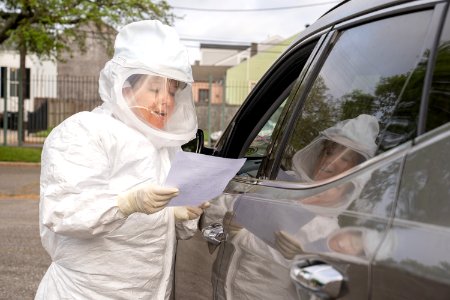
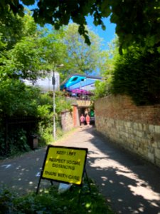
Armstrong Park photo
almost 3 years
Armstrong Park photo
almost 3 years
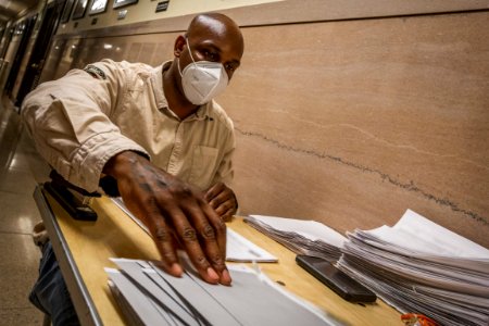
Delaware National Guard tests for COVID-19 at Dewey Beach photo
almost 3 years
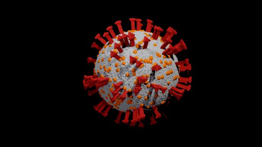
Coronavirus photo
almost 3 years

COVID-19 - The year of the virus photo
almost 3 years
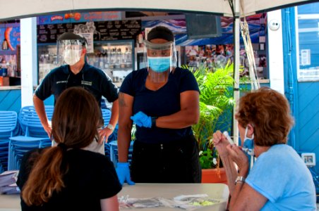
Respect photo
almost 3 years
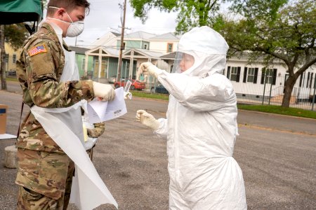
200323-Z-NI803-0053 photo
almost 3 years
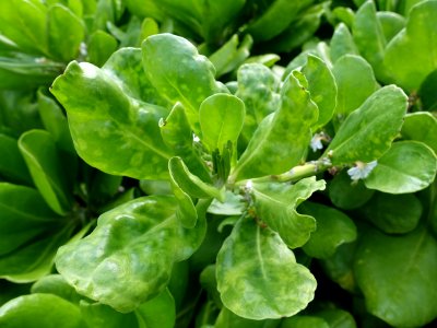
Beach naupaka (Scaevola taccada): Mosaic photo
almost 3 years
200722-Z-AL508-1045 photo
almost 3 years
200323-Z-NI803-0626 photo
almost 3 years

Protect yourself first, To be able to save people photo
almost 3 years
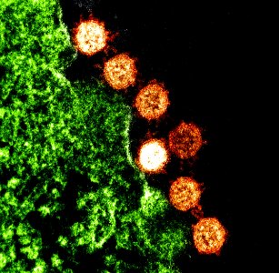
SARS Virus Particles photo
almost 3 years
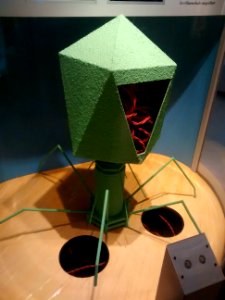
Virus (model) photo
almost 3 years

Hacking photo
almost 3 years
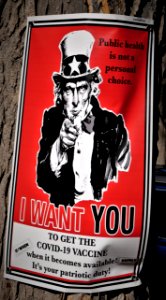
Uncle Sam COVID-19 Vaccine photo
almost 3 years
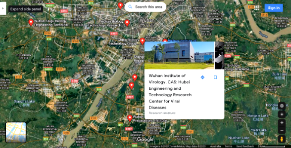
Wuhan photo
almost 3 years
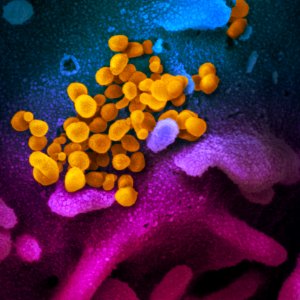
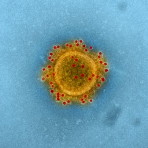
Novel Coronavirus SARS-CoV-2–This scanning electron microscope image shows SARS-CoV-2—also known as 2019-nCoV, the virus that causes COVID-19. Original image sourced from US Government department: The National Institute of Allergy and Infectious Diseases. Under US law this image is copyright free, please credit the government department whenever you can”. photo
about 3 years
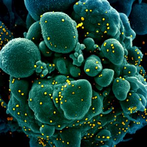
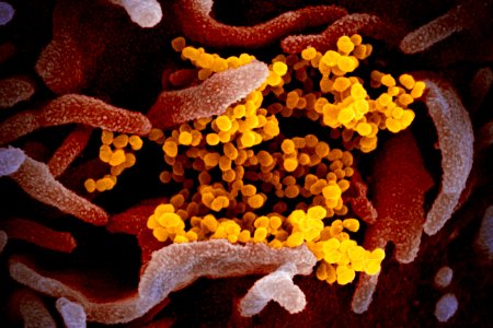
Novel Coronavirus SARS-CoV-2–Colorized scanning electron micrograph of an apoptotic cell (green) infected with SARS-COV-2 virus particles (yellow), isolated from a patient sample. Original image sourced from US Government department: The National Institute of Allergy and Infectious Diseases. Under US law this image is copyright free, please credit the government department whenever you can”. photo
about 3 years
Novel Coronavirus SARS-CoV-2–Colorized scanning electron micrograph of an apoptotic cell (blue) infected with SARS-COV-2 virus particles (red), isolated from a patient sample. Original image sourced from US Government department: The National Institute of Allergy and Infectious Diseases. Under US law this image is copyright free, please credit the government department whenever you can”. photo
about 3 years
MERS Coronavirus Particles–Colorized scanning electron micrograph of MERS virus particles (yellow) both budding and attached to the surface of infected VERO E6 cells (blue). Original image sourced from US Government department: The National Institute of Allergy and Infectious Diseases. Under US law this image is copyright free, please credit the government department whenever you can”. photo
about 3 years
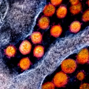
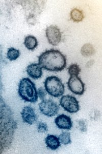
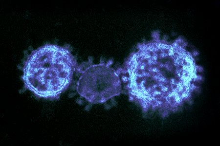
Novel Coronavirus SARS-CoV-2–Transmission electron micrograph of SARS-CoV-2 virus particles, isolated from a patient. Original image sourced from US Government department: The National Institute of Allergy and Infectious Diseases. Under US law this image is copyright free, please credit the government department whenever you can”. photo
about 3 years
Novel Coronavirus SARS-CoV-2–This scanning electron microscope image shows SARS-CoV-2 (yellow)—also known as 2019-nCoV, the virus that causes COVID-19. Original image sourced from US Government department: The National Institute of Allergy and Infectious Diseases. Under US law this image is copyright free, please credit the government department whenever you can”. photo
about 3 years
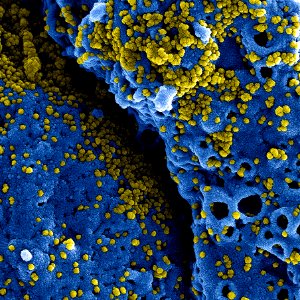
Novel Coronavirus SARS-CoV-2–Colorized scanning electron micrograph of an apoptotic cell (blue) heavily infected with SARS-COV-2 virus particles (yellow), isolated from a patient sample. Original image sourced from US Government department: The National Institute of Allergy and Infectious Diseases. Under US law this image is copyright free, please credit the government department whenever you can”. photo
about 3 years
MERS Coronavirus Particle–Middle East Respiratory Syndrome Coronavirus particle envelope proteins immunolabeled with Rabbit HCoV-EMC/2012 primary antibody and Goat anti-Rabbit 10 nm gold particles. Original image sourced from US Government department: The National Institute of Allergy and Infectious Diseases. Under US law this image is copyright free, please credit the government department whenever you can”. photo
about 3 years
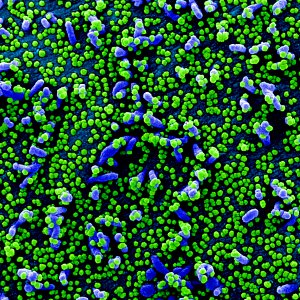
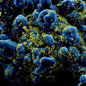
Novel Coronavirus SARS-CoV-2–Colorized scanning electron micrograph of a VERO E6 cell (blue) heavily infected with SARS-COV-2 virus particles (green), isolated from a patient sample. Original image sourced from US Government department: The National Institute of Allergy and Infectious Diseases. Under US law this image is copyright free, please credit the government department whenever you can”. photo
about 3 years
MERS Coronavirus Particles–Colorized transmission electron micrograph showing particles of the Middle East respiratory syndrome coronavirus that emerged in 2012. Original image sourced from US Government department: The National Institute of Allergy and Infectious Diseases. Under US law this image is copyright free, please credit the government department whenever you can”. photo
about 3 years
Novel Coronavirus SARS-CoV-2– This scanning electron microscope image shows SARS-CoV-2 (yellow)—also known as 2019-nCoV, the virus that causes COVID-19. Original image sourced from US Government department: The National Institute of Allergy and Infectious Diseases. Under US law this image is copyright free, please credit the government department whenever you can”. photo
about 3 years
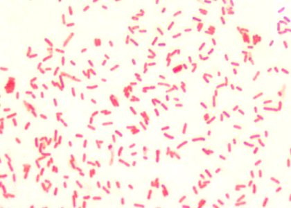
A 1000X photomicrograph magnification of numerous, rod-shaped, Gram–negative, Yersinia pestis bacilli. Original image sourced from US Government department: Public Health Image Library, Centers for Disease Control and Prevention. Under US law this image is copyright free, please credit the government department whenever you can”. photo
about 3 years
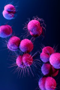
A medical illustration of drug–resistant, Neisseria gonorrhoeae bacteria. Original image sourced from US Government department: Public Health Image Library, Centers for Disease Control and Prevention. Under US law this image is copyright free, please credit the government department whenever you can”. photo
about 3 years
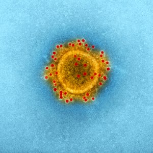
Transmission electron microscopic (TEM) image highlights the particle envelope of a single, spherical shaped, Middle East respiratory syndrome coronavirus (MERS-CoV) virion. Original image sourced from US Government department: Public Health Image Library, Centers for Disease Control and Prevention. Under US law this image is copyright free, please credit the government department whenever you can”. photo
about 3 years
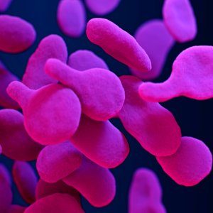
A medical illustration of drug–resistant, Mycoplasma genitalium bacteria. Original image sourced from US Government department: Public Health Image Library, Centers for Disease Control and Prevention. Under US law this image is copyright free, please credit the government department whenever you can”. photo
about 3 years
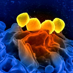
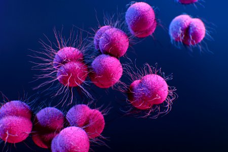
Group A Streptococcus (GAS), Streptococcus pyogenes bacteria, which were atop the surface of a human white blood cell (WBC), known as a neutrophil. Original image sourced from US Government department: Public Health Image Library, Centers for Disease Control and Prevention. Under US law this image is copyright free, please credit the government department whenever you can”. photo
about 3 years

A 3D illustration provides a graphical representation of a single norovirus virion, set against a beige background. Original image sourced from US Government department: Public Health Image Library, Centers for Disease Control and Prevention. Under US law this image is copyright free, please credit the government department whenever you can”. photo
about 3 years
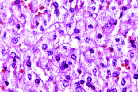
A 440X photomicrograph magnification of a hematoxylin and eosin (H&E)–stained liver tissue specimen, revealed the presence of cytoarchitectural changes indicative of fatty degeneration. Original image sourced from US Government department: Public Health Image Library, Centers for Disease Control and Prevention. Under US law this image is copyright free, please credit the government department whenever you can”. photo
about 3 years
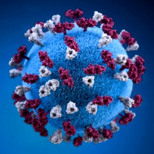
A 3D graphic representation of a spherical-shaped, measles virus particle, that was studded with glycoprotein tubercles. Original image sourced from US Government department: Public Health Image Library, Centers for Disease Control and Prevention. Under US law this image is copyright free, please credit the government department whenever you can”. photo
about 3 years
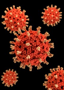
A 3D graphical representation of a number of Rotavirus virions, set against a black background. Original image sourced from US Government department: Public Health Image Library, Centers for Disease Control and Prevention. Under US law this image is copyright free, please credit the government department whenever you can”. photo
about 3 years
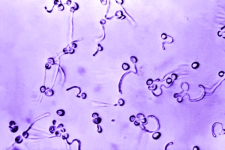
A photomicrograph of Candida albicans fungal spores. Original image sourced from US Government department: Public Health Image Library, Centers for Disease Control and Prevention. Under US law this image is copyright free, please credit the government department whenever you can”. photo
about 3 years
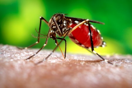
A female, Aedes aegypti mosquito obtaining a blood meal from a human host. Original image sourced from US Government department: Public Health Image Library, Centers for Disease Control and Prevention. Under US law this image is copyright free, please credit the government department whenever you can”. photo
about 3 years
A medical illustration of drug–resistant, Neisseria gonorrhoeae bacteria. Original image sourced from US Government department: Public Health Image Library, Centers for Disease Control and Prevention. Under US law this image is copyright free, please credit the government department whenever you can”. photo
about 3 years
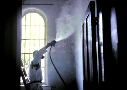
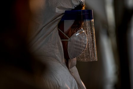
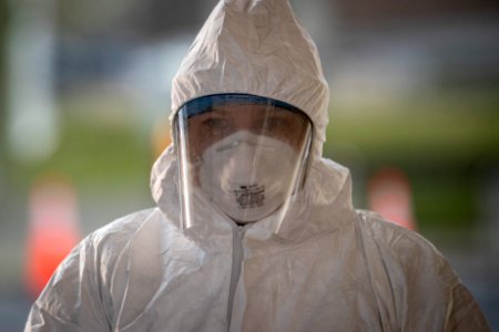
A worker wearing a protective mask and suit. Original image sourced from US Government department: Public Health Image Library, Centers for Disease Control and Prevention. Under US law this image is copyright free, please credit the government department whenever you can”. photo
about 3 years