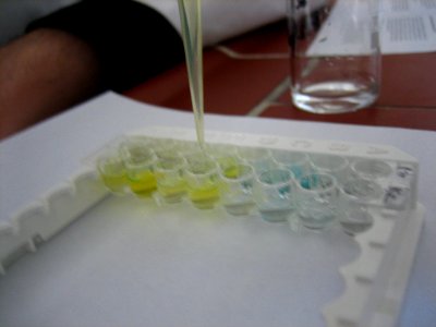
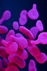
Something with proteines photo
almost 3 years
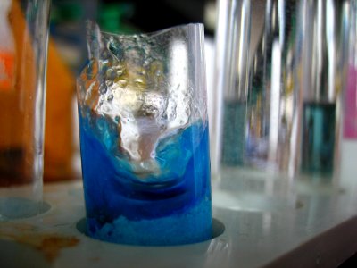
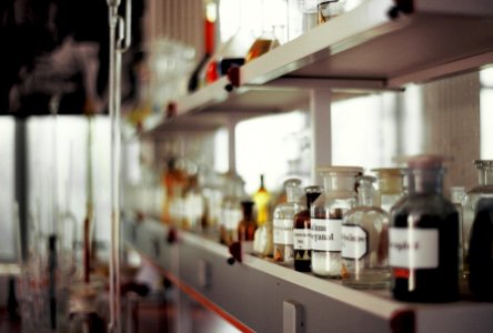
Chemicals in broken test tube photo
almost 3 years
201019-N-DA693-1002 photo
almost 3 years
let's cook photo
almost 3 years
im Kochstudio photo
almost 3 years
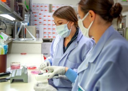
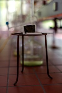
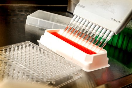
laboratory photo
almost 3 years

Rocket, Sztolnie Waliwskie w Walim, Polska photo
almost 3 years
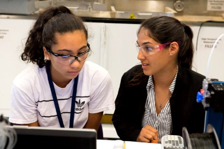
Army mentors students, educators in 3-D design and printing photo
almost 3 years
A medical illustration of drug–resistant, Mycoplasma genitalium bacteria. Original image sourced from US Government department: Public Health Image Library, Centers for Disease Control and Prevention. Under US law this image is copyright free, please credit the government department whenever you can”. photo
about 3 years
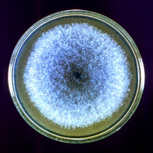
A Petri dish culture plate containing a growth medium of Sabouraud dextrose agar, which had been inoculated with the fungal organism from the genus, Syncephalastrum, strain A-423. Original image sourced from US Government department: Public Health Image Library, Centers for Disease Control and Prevention. Under US law this image is copyright free, please credit the government department whenever you can”. photo
about 3 years
A scientist testing bacterial samples for their antimicrobial susceptibility. Original image sourced from US Government department: Public Health Image Library, Centers for Disease Control and Prevention. Under US law this image is copyright free, please credit the government department whenever you can”. photo
about 3 years
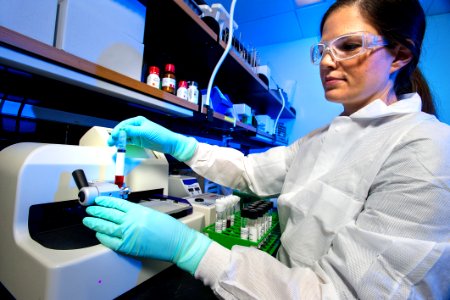
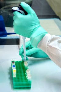
Scientist pipetting samples of bronchoalveolar lavage (BAL) fluid. Original image sourced from US Government department: Public Health Image Library, Centers for Disease Control and Prevention. Under US law this image is copyright free, please credit the government department whenever you can”. photo
about 3 years
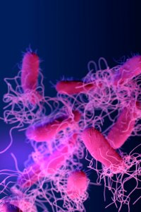
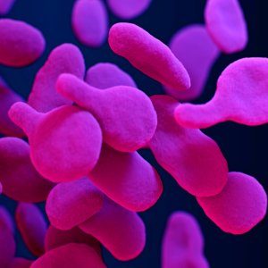
A medical illustration of drug–resistant, nontyphoidal, Salmonella sp. bacteria. Original image sourced from US Government department: Public Health Image Library, Centers for Disease Control and Prevention. Under US law this image is copyright free, please credit the government department whenever you can”. photo
about 3 years
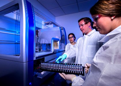
Scientists using automated methods to extract bacterial DNA, for use in whole genome sequencing (WGS). Original image sourced from US Government department: Public Health Image Library, Centers for Disease Control and Prevention. Under US law this image is copyright free, please credit the government department whenever you can”. photo
about 3 years
Laboratory technician working with electronic cigarettes. Original image sourced from US Government department: Public Health Image Library, Centers for Disease Control and Prevention. Under US law this image is copyright free, please credit the government department whenever you can”. photo
about 3 years
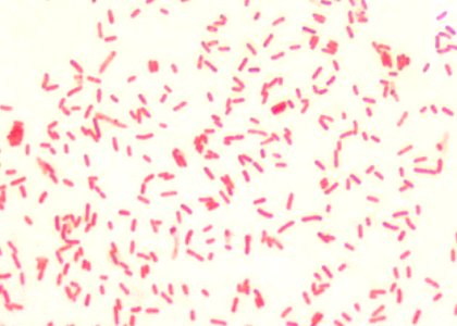
A 1000X photomicrograph magnification of numerous, rod-shaped, Gram–negative, Yersinia pestis bacilli. Original image sourced from US Government department: Public Health Image Library, Centers for Disease Control and Prevention. Under US law this image is copyright free, please credit the government department whenever you can”. photo
about 3 years
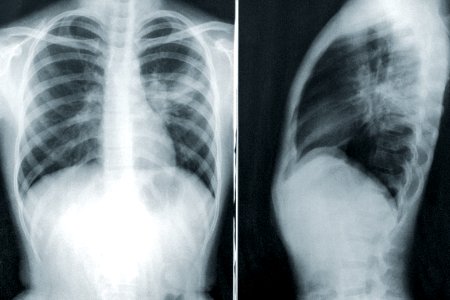
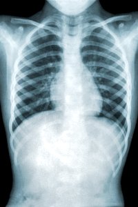
Two chest x-rays in the case of a child with a case of mycoplasma pneumonia. Original image sourced from US Government department: Public Health Image Library, Centers for Disease Control and Prevention. Under US law this image is copyright free, please credit the government department whenever you can”. photo
about 3 years
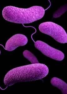
A 3D image of a number of oblong–shaped, Vibrio parahaemolyticus bacteria Original image sourced from US Government department: Public Health Image Library, Centers for Disease Control and Prevention. Under US law this image is copyright free, please credit the government department whenever you can”. photo
about 3 years

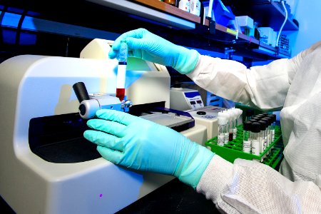
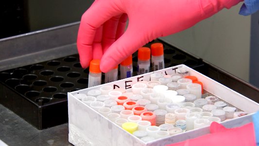
A test tube tray containing a number of purple tipped vacutainer tubes. Original image sourced from US Government department: Public Health Image Library, Centers for Disease Control and Prevention. Under US law this image is copyright free, please credit the government department whenever you can”. photo
about 3 years
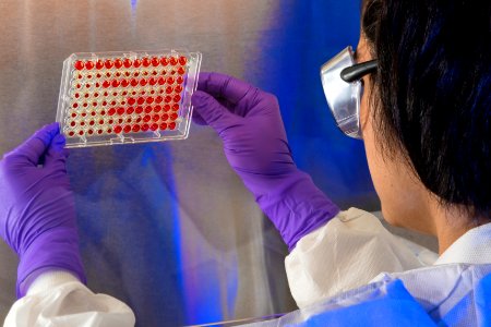
A scientist examining the results of a hemagglutinin inhibition (HI) test. Original image sourced from US Government department: Public Health Image Library, Centers for Disease Control and Prevention. Under US law this image is copyright free, please credit the government department whenever you can”. photo
about 3 years
A medical illustration of drug–resistant, Mycoplasma genitalium bacteria. Original image sourced from US Government department: Public Health Image Library, Centers for Disease Control and Prevention. Under US law this image is copyright free, please credit the government department whenever you can”. photo
about 3 years
A scientist adding a stool sample to the cell culture medium, along with glass beads. Original image sourced from US Government department: Public Health Image Library, Centers for Disease Control and Prevention. Under US law this image is copyright free, please credit the government department whenever you can”. photo
about 3 years
A scientist holding bacterial samples for their antimicrobial susceptibility. Original image sourced from US Government department: Public Health Image Library, Centers for Disease Control and Prevention. Under US law this image is copyright free, please credit the government department whenever you can”. photo
about 3 years
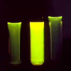
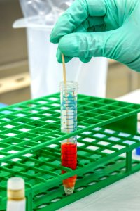
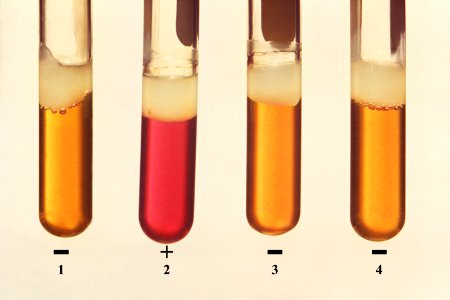
Three test tubes used in a Legionella pneumophila bacteria detection test. Original image sourced from US Government department: Public Health Image Library, Centers for Disease Control and Prevention. Under US law this image is copyright free, please credit the government department whenever you can”. photo
about 3 years
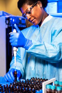
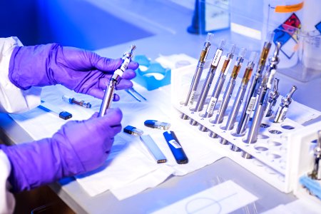
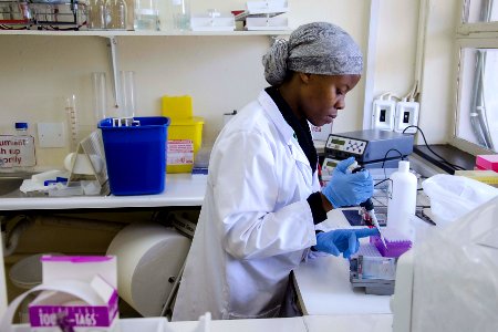
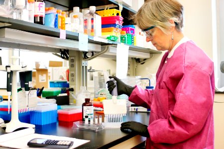
A scientist implementing molecular testing. Original image sourced from US Government department: Public Health Image Library, Centers for Disease Control and Prevention. Under US law this image is copyright free, please credit the government department whenever you can”. photo
about 3 years
A scientist implementing molecular testing. Original image sourced from US Government department: Public Health Image Library, Centers for Disease Control and Prevention. Under US law this image is copyright free, please credit the government department whenever you can”. photo
about 3 years
A Petri dish culture plate, which had contained a growth medium of sheep blood agar (SBA), and which was inoculated with Gram–positive, Burkholderia pseudomallei bacteria. Original image sourced from US Government department: Public Health Image Library, Centers for Disease Control and Prevention. Under US law this image is copyright free, please credit the government department whenever you can”. photo
about 3 years
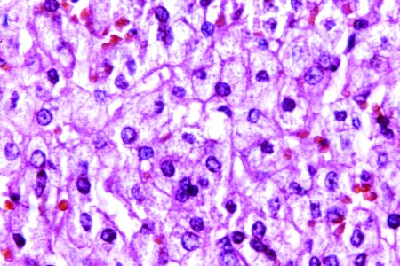
A 440X photomicrograph magnification of a hematoxylin and eosin (H&E)–stained liver tissue specimen, revealed the presence of cytoarchitectural changes indicative of fatty degeneration. Original image sourced from US Government department: Public Health Image Library, Centers for Disease Control and Prevention. Under US law this image is copyright free, please credit the government department whenever you can”. photo
about 3 years
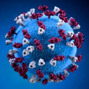
A 3D graphic representation of a spherical-shaped, measles virus particle, that was studded with glycoprotein tubercles. Original image sourced from US Government department: Public Health Image Library, Centers for Disease Control and Prevention. Under US law this image is copyright free, please credit the government department whenever you can”. photo
about 3 years
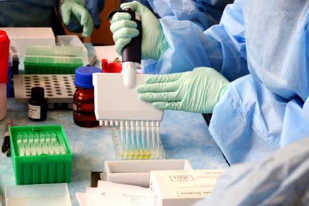
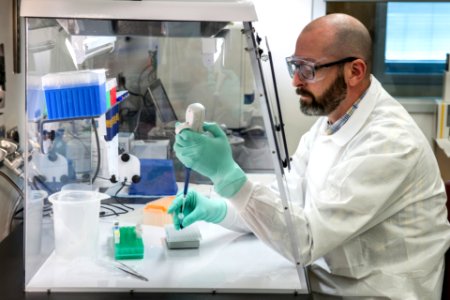
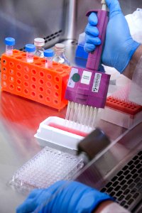
Lab technician adding a solution to an Enzyme–Linked Immunosorbent Assay (ELISA) plate Original image sourced from US Government department: Public Health Image Library, Centers for Disease Control and Prevention. Under US law this image is copyright free, please credit the government department whenever you can”. photo
about 3 years

A scientist holding a culture test plate. Original image sourced from US Government department: Public Health Image Library, Centers for Disease Control and Prevention. Under US law this image is copyright free, please credit the government department whenever you can”. photo
about 3 years
Four test tubes of bacterial microorganism. Original image sourced from US Government department: Public Health Image Library, Centers for Disease Control and Prevention. Under US law this image is copyright free, please credit the government department whenever you can”. photo
about 3 years
Chest x-ray of a patient with mycoplasma pneumonia. Original image sourced from US Government department: Public Health Image Library, Centers for Disease Control and Prevention. Under US law this image is copyright free, please credit the government department whenever you can”. photo
about 3 years
Scientist testing with virus through test tubes. Original image sourced from US Government department: Public Health Image Library, Centers for Disease Control and Prevention. Under US law this image is copyright free, please credit the government department whenever you can”. photo
about 3 years
Scientist testing with virus through test tubes. Original image sourced from US Government department: Public Health Image Library, Centers for Disease Control and Prevention. Under US law this image is copyright free, please credit the government department whenever you can”. photo
about 3 years
Test Tubes from experimenting on coronavirus. Original image sourced from US Government department: Public Health Image Library, Centers for Disease Control and Prevention. Under US law this image is copyright free, please credit the government department whenever you can”. photo
about 3 years
Microbiologist preparing for antibody testing. A healthcare provider donning a pair of green latex gloves in order to protect herself during her subsequent interaction her next patient. Original image sourced from US Government department: Public Health Image Library, Centers for Disease Control and Prevention. Under US law this image is copyright free, please credit the government department whenever you can”. photo
about 3 years
Sample of test tubes. A healthcare provider donning a pair of green latex gloves in order to protect herself during her subsequent interaction her next patient. Original image sourced from US Government department: Public Health Image Library, Centers for Disease Control and Prevention. Under US law this image is copyright free, please credit the government department whenever you can”. photo
about 3 years
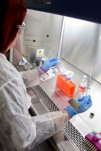
Laboratory technician working with a cell culture solution, using a syringe to add some of the sample to a glass bottle. A healthcare provider donning a pair of green latex gloves in order to protect herself during her subsequent interaction her next patient. Original image sourced from US Government department: Public Health Image Library, Centers for Disease Control and Prevention. Under US law this image is copyright free, please credit the government department whenever you can”. photo
about 3 years
Test Tubes from experimenting on coronavirus. Original image sourced from US Government department: Public Health Image Library, Centers for Disease Control and Prevention. Under US law this image is copyright free, please credit the government department whenever you can”. photo
about 3 years