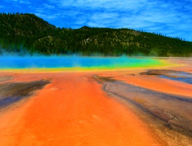
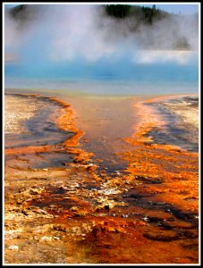
Yellowstone national park, United states, Bacteria photo
téměř 3 roky
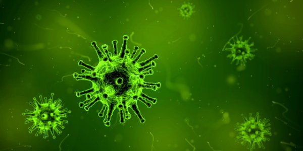
Virus image photo
téměř 3 roky
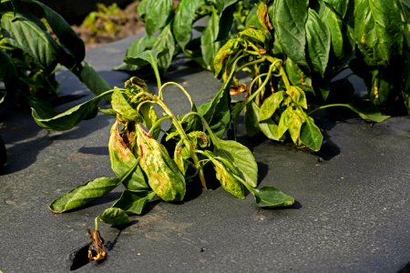
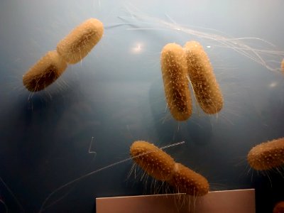
20170106-ARS-STA-0002 photo
téměř 3 roky

20170106-ARS-STA-0001 photo
téměř 3 roky

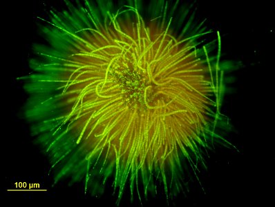
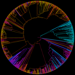
Color growth from virtual bacteria (generative) photo
téměř 3 roky
Green in Your Life photo
téměř 3 roky
Harmful Water Organisms photo
téměř 3 roky
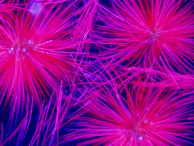
Mineral Pond, Pool of Life, Yellowstone National Park, Wyoming photo
téměř 3 roky
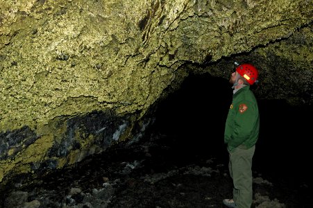
Bacteria growing within water drops colors the walls golden in Golden Dome Cave in Lava Beds National Monument. photo
téměř 3 roky
HUGE-B 500-NCBI PGT photo
téměř 3 roky
Bacteria photo
téměř 3 roky
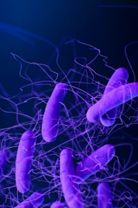
A medical illustration of Clostridioides difficile bacteria, formerly known as Clostridium difficile. Original image sourced from US Government department: Public Health Image Library, Centers for Disease Control and Prevention. Under US law this image is copyright free, please credit the government department whenever you can”. photo
asi 3 roky
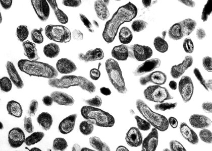
Transmission electron microscopic (TEM) image, revealed ultrastructural details exhibited by numerous, Coxiella burnetii bacteria, which cause the worldwide disease known, as Q fever. Original image sourced from US Government department: Public Health Image Library, Centers for Disease Control and Prevention. Under US law this image is copyright free, please credit the government department whenever you can”. photo
asi 3 roky
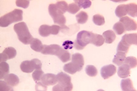
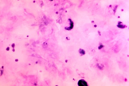
A 1125X photomicrograph magnification of a Giemsa stained, thin film blood smear, revealed a mature, Plasmodium malariae schizont. Original image sourced from US Government department: Public Health Image Library, Centers for Disease Control and Prevention. Under US law this image is copyright free, please credit the government department whenever you can”. photo
asi 3 roky
A medical illustration of drug–resistant, Mycoplasma genitalium bacteria. Original image sourced from US Government department: Public Health Image Library, Centers for Disease Control and Prevention. Under US law this image is copyright free, please credit the government department whenever you can”. photo
asi 3 roky
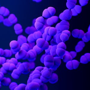
A medical illustration of drug–resistant, Streptococcus pneumoniae bacteria. Original image sourced from US Government department: Public Health Image Library, Centers for Disease Control and Prevention. Under US law this image is copyright free, please credit the government department whenever you can”. photo
asi 3 roky
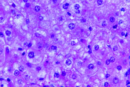
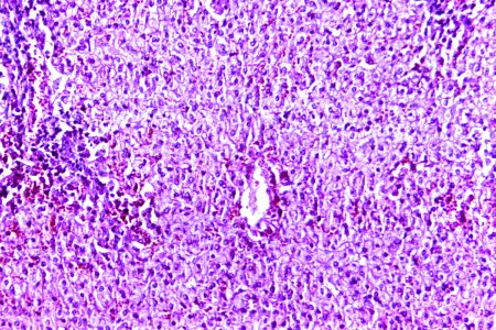
A 440X photomicrograph magnification of a hematoxylin and eosin (H&E)–stained liver tissue specimen, revealed the presence of cytoarchitectural changes indicative of fatty degeneration. Original image sourced from US Government department: Public Health Image Library, Centers for Disease Control and Prevention. Under US law this image is copyright free, please credit the government department whenever you can”. photo
asi 3 roky
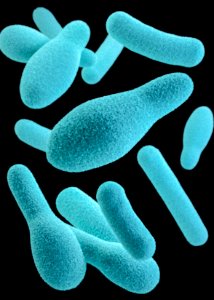
A 3D image of a group of anaerobic, spore-forming, Clostridium sp. organisms. Original image sourced from US Government department: Public Health Image Library, Centers for Disease Control and Prevention. Under US law this image is copyright free, please credit the government department whenever you can”. photo
asi 3 roky
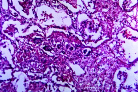
A 125X photomicrograph magnification of a hematoxylin and eosin (H&E)–stained lung tissue specimen, revealed the presence of cytoarchitectural changes indicative of a blastomycosis fungal infection. Original image sourced from US Government department: Public Health Image Library, Centers for Disease Control and Prevention. Under US law this image is copyright free, please credit the government department whenever you can”. photo
asi 3 roky
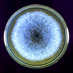
A Petri dish culture plate containing a growth medium of Sabouraud dextrose agar, which had been inoculated with the fungal organism from the genus, Syncephalastrum, strain A-423. Original image sourced from US Government department: Public Health Image Library, Centers for Disease Control and Prevention. Under US law this image is copyright free, please credit the government department whenever you can”. photo
asi 3 roky
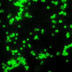
A photomicrograph of a direct fluorescent antibody (DFA)–stained specimen, revealing the presence of numerous Francisella tularensis coccobacilli. Original image sourced from US Government department: Public Health Image Library, Centers for Disease Control and Prevention. Under US law this image is copyright free, please credit the government department whenever you can”. photo
asi 3 roky

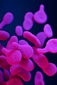
A medical illustration of Clostridioides difficile bacteria, formerly known as Clostridium difficile. Original image sourced from US Government department: Public Health Image Library, Centers for Disease Control and Prevention. Under US law this image is copyright free, please credit the government department whenever you can”. photo
asi 3 roky
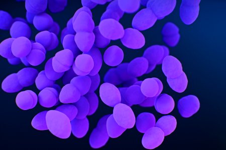
A medical illustration of vancomycin–resistant, Enterococci sp. bacteria. Original image sourced from US Government department: Public Health Image Library, Centers for Disease Control and Prevention. Under US law this image is copyright free, please credit the government department whenever you can”. photo
asi 3 roky
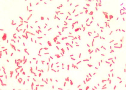
A photomicrograph reveals numerous Gram–negative, Francisella tularensis coccobacilli, the bacterium responsible for causing the disease, tularemia. Original image sourced from US Government department: Public Health Image Library, Centers for Disease Control and Prevention. Under US law this image is copyright free, please credit the government department whenever you can”. photo
asi 3 roky
A 1125X photomicrograph magnification of Giemsa stained blood smear, revealed a number of Plasmodium falciparum parasites, in the form of gametocytes, and ring-form trophozoites. Original image sourced from US Government department: Public Health Image Library, Centers for Disease Control and Prevention. Under US law this image is copyright free, please credit the government department whenever you can”. photo
asi 3 roky
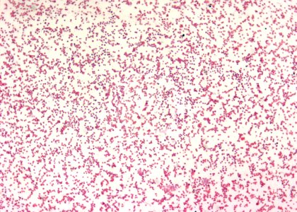
A 100X photomicrograph magnification of a malachite green spore stained sample, revealed the presence of numerous Bacillus sp. bacteria. Original image sourced from US Government department: Public Health Image Library, Centers for Disease Control and Prevention. Under US law this image is copyright free, please credit the government department whenever you can”. photo
asi 3 roky
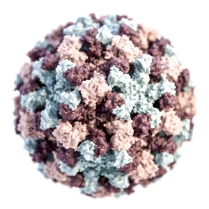
A 3D illustration provides a graphical representation of a single norovirus virion, set against a white background. Original image sourced from US Government department: Public Health Image Library, Centers for Disease Control and Prevention. Under US law this image is copyright free, please credit the government department whenever you can”. photo
asi 3 roky
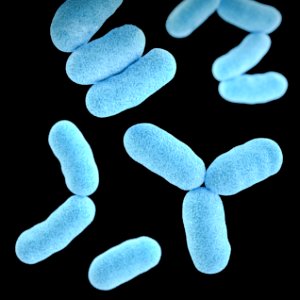
A 3D image of a group of Gram–positive, Corynebacterium diphtheriae, bacteria. Original image sourced from US Government department: Public Health Image Library, Centers for Disease Control and Prevention. Under US law this image is copyright free, please credit the government department whenever you can”. photo
asi 3 roky
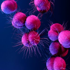
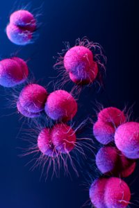
A medical illustration of drug–resistant, Neisseria gonorrhoeae bacteria. Original image sourced from US Government department: Public Health Image Library, Centers for Disease Control and Prevention. Under US law this image is copyright free, please credit the government department whenever you can”. photo
asi 3 roky
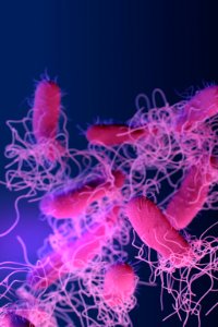
A medical illustration of drug–resistant, nontyphoidal, Salmonella sp. bacteria. Original image sourced from US Government department: Public Health Image Library, Centers for Disease Control and Prevention. Under US law this image is copyright free, please credit the government department whenever you can”. photo
asi 3 roky

A medical illustration of vancomycin–resistant, Enterococci sp. bacteria. Original image sourced from US Government department: Public Health Image Library, Centers for Disease Control and Prevention. Under US law this image is copyright free, please credit the government department whenever you can”. photo
asi 3 roky
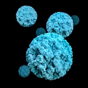
A 3D graphical representation of a number of norovirus virions, set against a black background. Original image sourced from US Government department: Public Health Image Library, Centers for Disease Control and Prevention. Under US law this image is copyright free, please credit the government department whenever you can”. photo
asi 3 roky
A 100X photomicrograph magnification of photomicrograph of a hematoxylin and eosin (H&E) stained liver tissue specimen, revealed the presence of histopathologic changes indicative of fatty degeneration. Original image sourced from US Government department: Public Health Image Library, Centers for Disease Control and Prevention. Under US law this image is copyright free, please credit the government department whenever you can”. photo
asi 3 roky
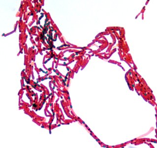
A 1000X photomicrograph magnification of numerous, rod-shaped, Gram–negative, Yersinia pestis bacilli. Original image sourced from US Government department: Public Health Image Library, Centers for Disease Control and Prevention. Under US law this image is copyright free, please credit the government department whenever you can”. photo
asi 3 roky
A medical illustration of drug–resistant, Neisseria gonorrhoeae bacteria. Original image sourced from US Government department: Public Health Image Library, Centers for Disease Control and Prevention. Under US law this image is copyright free, please credit the government department whenever you can”. photo
asi 3 roky

A medical illustration of clindamycin–resistant group B Streptococcus bacteria. Original image sourced from US Government department: Public Health Image Library, Centers for Disease Control and Prevention. Under US law this image is copyright free, please credit the government department whenever you can”. photo
asi 3 roky
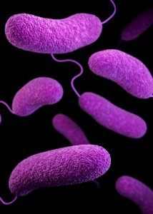
A 3D image of a number of oblong–shaped, Vibrio parahaemolyticus bacteria Original image sourced from US Government department: Public Health Image Library, Centers for Disease Control and Prevention. Under US law this image is copyright free, please credit the government department whenever you can”. photo
asi 3 roky
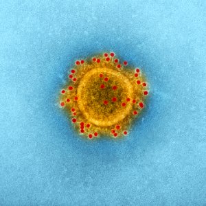
Transmission electron microscopic (TEM) image highlights the particle envelope of a single, spherical shaped, Middle East respiratory syndrome coronavirus (MERS-CoV) virion. Original image sourced from US Government department: Public Health Image Library, Centers for Disease Control and Prevention. Under US law this image is copyright free, please credit the government department whenever you can”. photo
asi 3 roky
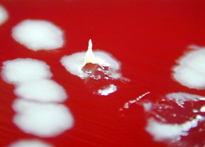
A petri dish culture plate contained a growth medium of sheep blood agar (SBA), which was inoculated with Bacillus anthracis bacteria. Original image sourced from US Government department: Public Health Image Library, Centers for Disease Control and Prevention. Under US law this image is copyright free, please credit the government department whenever you can”. photo
asi 3 roky
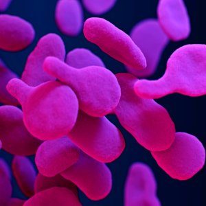
A medical illustration of drug–resistant, Mycoplasma genitalium bacteria. Original image sourced from US Government department: Public Health Image Library, Centers for Disease Control and Prevention. Under US law this image is copyright free, please credit the government department whenever you can”. photo
asi 3 roky